Cephalometric landmarks
Main sources of uncertainty in the landmarking of cephalometric points
Accurately locating the cephalometric landmarks when marking a photograph can prove to be an incredibly challenging task. The main sources of variability are:
a) Variability in the light and shadow distribution of the image.
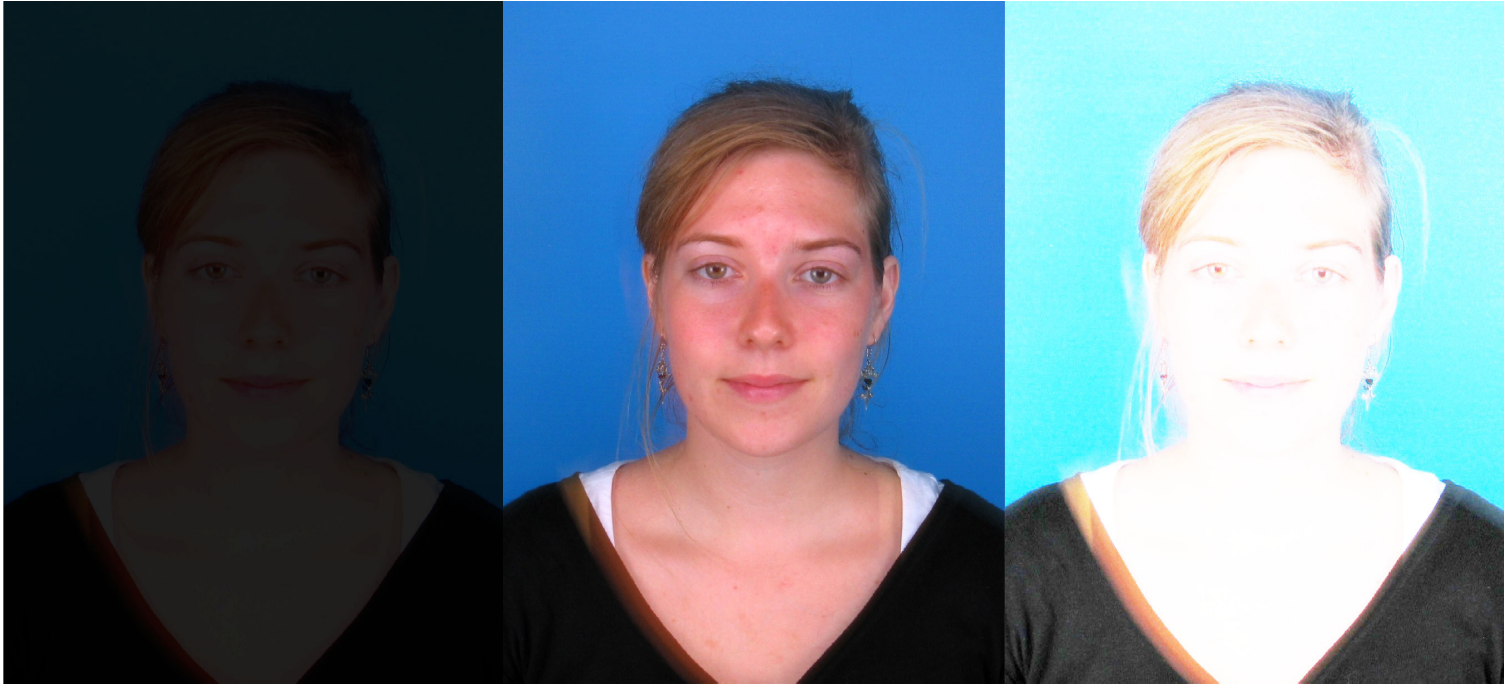 Figure 1. Examples of an underexposed photograph (left), a photograph with good exposure (centre) and an overexposed photograph (right). Notice the loss of details in the under and overexposed areas.
b) Subpar focus of the camera, preventing from a precise appraisal of the morphological features.
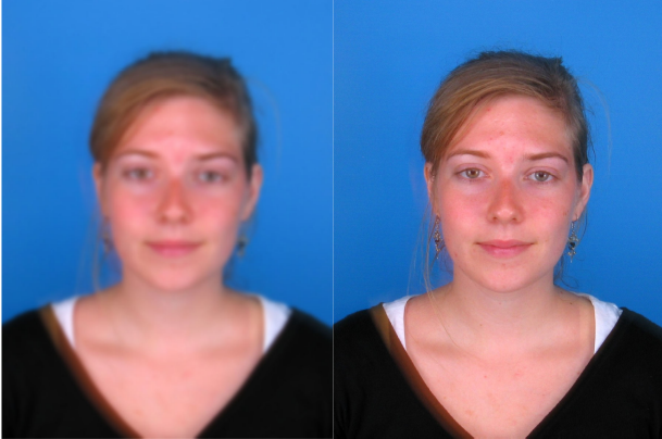 Figure 2. Example of an out of focus photograph (left) and a focused photograph (right).
c) Low resolution of the image.
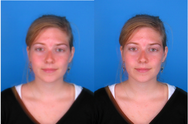 Figure 3. Examples of a low-resolution photograph (left) and a high/standard resolution photograph (right).
d) Face pose in the photograph (lateral, frontal or oblique) and facial expressions.
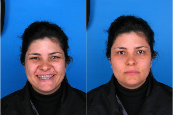 Figure 4. Example of the distortion caused by facial expression, same subject smiling (left) and with a neutral expression (right).
e) Presence of obscuring objects that preclude the view of facial structures (such as glasses, hair, beards, hats, etc.).
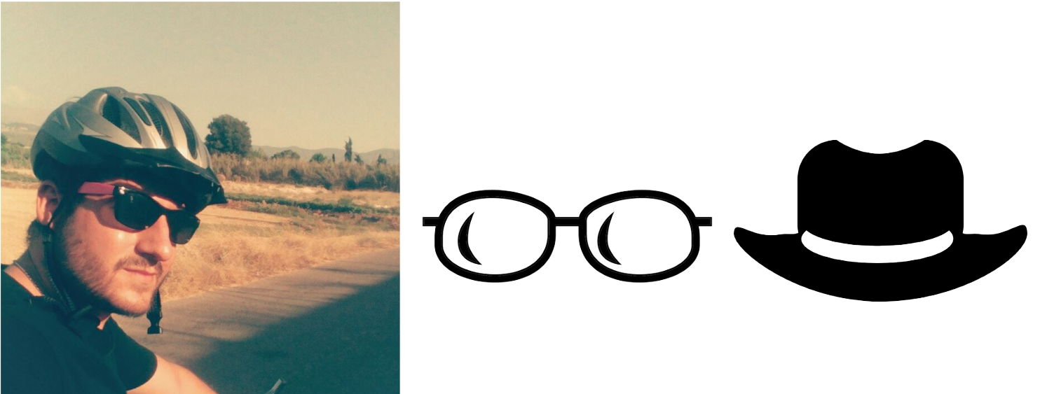 Figure 5. Obscuring objects (helmet and sunglasses) that hamper the cephalometric landmarking process.
Recommendations for cephalometric landmarking
a) For optimal marking on frontal photographs, a resolution of at least 180 pixels in the width of the head, or approximately 90 pixels between the pupils (ISO International Standard ISO / IEC JTC 1 / SC 37 N506) is advised.
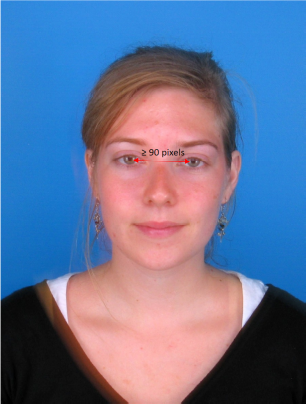 Figure 6. Recommended interpupillary distance.
b) In lateral face pose photographs (left or right) the contralateral point of bilateral landmarks cannot be estimated (for example, left and right exocanthion).
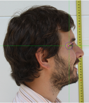 Figure 7. Lateral view image (right profile). The landmarks in the left profile cannot be estimated.
c) When placing bilateral facial landmarks, the anatomical laterality of the subject and not that of the observer should be considered.
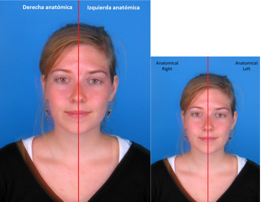 Figure 8. Anatomical laterality in a photograph.
Types of cephalometric landmarks
The accurate positioning of facial points is key before any facial analysis. More so, when it is done on photographs that may be the only available AM data or play an important role in a forensic evaluation. It should be noted that the precision in the location varies among landmarks (Campomanes-Álvarez et al. 2015).
Following Bookstein (1991), three types of landmarks can be discerned:
- Type 1: those where three structures meet (e.g. suture joints, nasion, subnasale o endocanthion).
- Type 2: those located on points of maximum curvature or other morphogenetic processes (e.g. exocanthion or labiale superius), generally with biomechanical implications, such as the tips of bony processes for muscle attachments in craniometric landmarks.
- Type 3: those located along a morphometric curve or surface, e.g. alare, gonion or zygion.
Nomenclature and definitions of the main cephalometric landmarks for Craniofacial Superimposition
Median landmarks:
- Vertex (v´): Most superior point of the head.
- Trichion (tr´): Midpoint of the hairline; determined on a widow’s peak as the projection through the midline from both sides.
- Glabella (g´): Most anterior midline point on the forehead, in the region of the superciliary ridges.
- Nasion (n´): Point directly anterior to the nasofrontal suture, in the midline, overlying n,.
- Subnasale (sn´): Median point at the junction between the lower border of the nasal septum and the philtrum area.
- Labiale superius (ls´): Midpoint of the vermillion border of the upper lip (not identical and not to be confused for Labrale superius).
- Stomion (sto´): Midline point of the labial fissure when the lips are closed naturally, with teeth shut in the natural position; if not in the midline, then below the philtrum.
- Labiale inferius (li´): Midpoint of the vermillion border of the liwer lip (identical to labrale inferius).
- Supramentale (sm´): Deepest midline point of the mentolabial sulcus.
- Pogonion (pg´): Most anterior midpoint of the chin, located on the skin surface anterior to the identical bony landmark of the mandible.
- Gnathion (gn´): Median point halfway between pg´and me´.
- Menton (me´): Most inferior median point of the chin.
Bilateral landmarks:
- Frontotemporale (ft´): Point of concavity on each side of the forehead above the supraorbital rim, lateral to the elevation of the linea temporalis.
- Mid-supraorbital (mso´): Point anteriorly adjacent to the supraorbital rim, at a line that vertically bisects the orbit.
- Frontozygomaticus (fz´): Most lateral point on the frontozygomatic suture, identified by palpation of the suture line at the superolateral corner of the orbit.
- Exocanthion (ec´): Most lateral point of the palpebral fissure, at the outer comissure of the eye; best seen when subject is gazing upward.
- Endocanthion (en´): Most medial point of the palpebral fissure, at the inner comissure of the eye; best seen when subject is gazing upward.
- Tragion (t´): Located at the notch above the tragus of the ear (the cartilaginous projection anterior to the external auditory canal), where the upper edge of the cartilage disappears into the skin of the face.
- Zygion (zy´): Most lateralpoint overlying each zygomatic arch, identified as the point of máximum bizygomatic breadth of the face.
- Alare (al´): The most lateral point on the nasal ala.
- Cheilion (ch´): Outer corners of the mouth where the outer edges of the upper and lower vermilion meet.
- Gonion (go´): Most lateral point on the mandibular angle, adjacent to go, identified by palpation.
Dispersion in the location of cephalometric landmarks
The quantification of the error in the location of facial landmarks on photographs has been researched by Cummaudo et al. (2013). In the cited work the inter and intraobserver dispersion on two photographs of the same subject in frontal and lateral pose and on eight photographs of different individuals of both sexes. In the interobserver study, 24 researchers located 18 facial landmarks 20 times with 24 hours intervals. The results showed the most significant dispersion in frontal photographs presented in landmarks like gonion, zygion and frontotemporale; while lateral photographs presented the most dispersion in gnathion, pogonion and tragion. On the other hand, the least dispersion in frontal photographs presented in endocanthion and stomion; and subnasale in lateral photographs. However, this study failed to assess the precision in landmark location in statistical significance terms.
Researchers from the University of Granada (Campomanes-Álvarez et al., 2015), conducted a study following this line, regarding the precision in the location of facial landmarks in 2D facial images, including statistical analysis and considering the sources of uncertainty such as landmark type, face pose, inter and intraobserver dispersion and the observer experience. For the interobserver study, 18 landmarks were used in 3 different poses (frontal, lateral and oblique view) and two types of observers (experts and students). For the intraobserver study the dispersion in 13 landmarks marked in 5 photographs in frontal and oblique view was assessed. Three different observers located the landmarks in 24 hours intervals. The results of the study showed that:
- Concerning interobserver dispersion, gnathion, gonion and zygion (Type 3 landmarks) were statistically significant, but their effect in dispersion was influenced by the type of image. Gonion and zygion had statistically significant dispersion in frontal images. The greatest dispersion was observed in gonion, zigion and gnathion in oblique view. In lateral photographs, the dispersion was greater in gonion, while zygion presented the most dispersion in all the 3 views.
- Concerning the frequency analysis, glabella, nasion, subnasale, labiale superius and pogonion were the most regularly located landmarks in the three types of images, marked in at least 80% of the cases. Conversely, the least frequently marked were labiale inferius and mentón, with a 50 and 25% respectively. Both endocanthion, exocanthion, and alare were placed in over 70% of frontal and oblique photographs. Both gonion presented the most significant difference in location among the 3 vies. Generally, Type 1 and 2 landmarks are located more often than Type 3 ones.
- The intraobserver analysis revealed that zygion and gonion were significantly more challenging to locate than other facial landmarks. The considerable variability when placing these landmarks can be because they are more difficult to locate without palpation. Overall, intraobserver dispersion was greater in oblique images than in frontal and lateral ones.
- Some type 3 landmarks tend to vary greatly when it comes to accurately determining their location. They are associated with a high error rate, statistically significant both at inter and intraobserver levels.