Facial areas
A review of the facial areas and their anatomy is necessary to ease the description of cephalometric landmarks and the understanding of the landmarking process – the anatomical structures in this area will be referenced. According to George (2007), the face can be divided into 8 areas:
- Frontal area
- Nasal area
- Labial area
- Mental area
- Orbital area
- Zygomaxillary area
- Buccomandibular area
- Auricular area
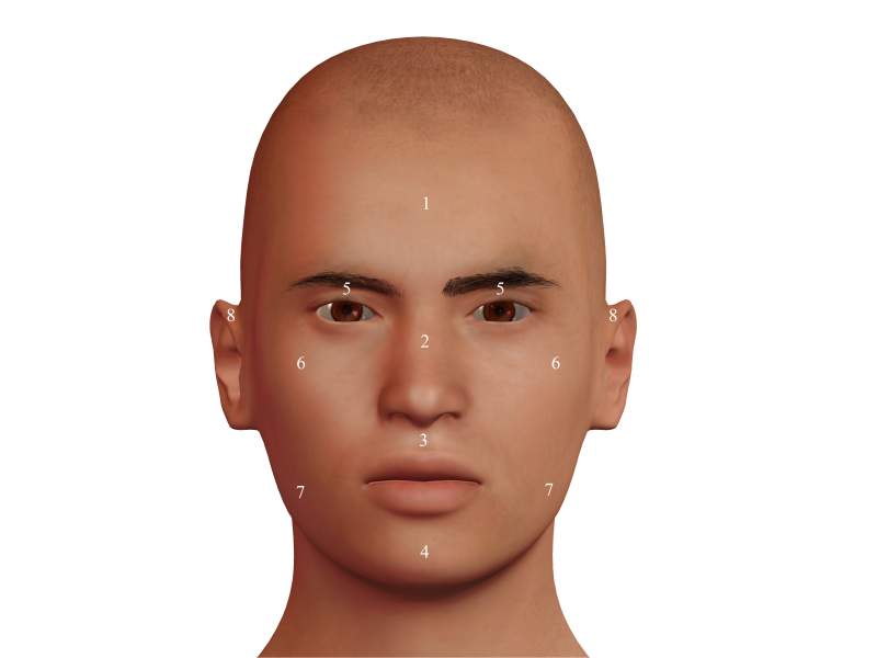 Figure 1. Facial areas: 1 = Frontal; 2 = Nasal; 3 = Labial; 4 = Mental; 5 = Orbital; 6 = Zygomaxilar; 7 = Bucomandibular; 8 = Auricular.
Below, the main facial structures in the frontal area, the eye, nose, mouse and ear, will be described according to George (2007) definitions.
Orbital area
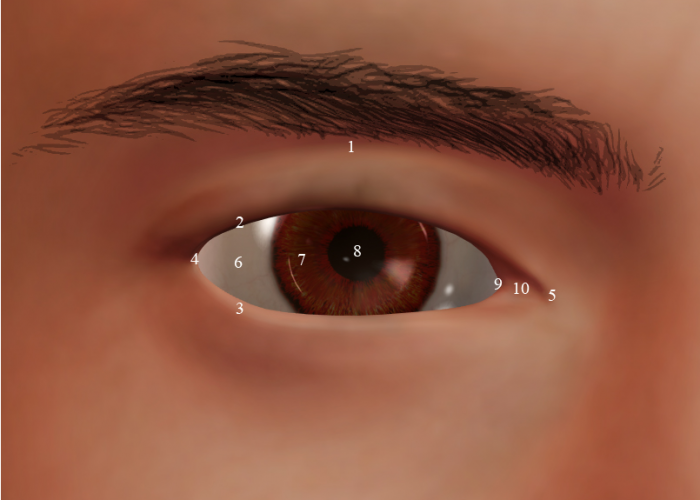 Figure 2. Orbital area: 1 = Superior orbital sulcus; 2 = Free margin of the upper eyelid; 3 = Free margin of the lower eyelid; 4 = Lateral canthus; 5 = Medial canthus; 6 = Cornea / sclera; 7 = Iris; 8= Pupil; 9 = Plica semilunaris; 10 = Lacrimal caruncle.
- Superior orbital (palpebral) sulcus/fissure: the fold between the upper eyelid and the skin over the upper orbital septum; a line connecting the left and right sulcus through the nasion in the medial sagittal plane; this skin tends to sag with aging, hiding the upper orbital eyelid, which should be taken into consideration on subjects over the age of 50-.
- Free margin of the upper eyelid (palpebrae superioris): the upper eyelid is lifted by the levator palpebrae superioris muscle, located inside the orbit. This muscle is joint to the tarsal plate, a fibrous, dense plate that conforms the “skeleton of the eyelid”. This is one of the extra ocular muscles and not linked to facial expresión development.
- Free margin of the lower eyelid (palpebrae inferioris): this eyelid also has a tarsal plate, but no independent muscle.
- Lateral canthus: the lateral corner of the eye (the cephalometric exocanthion).
- Medial canthus: the medial corner of the eye (the cephalometric endocanthion).
- Cornea / sclera: the external fibrous film of the eyeball; the sclera is opaque (the white of the eye) and the cornea is the clear portion right on top of the iris and pupil.
- Iris: it is a pigmented disc that contains two dispositions of smooth muscle tissue tasked with regulating the diameter of the pupil; the iris dilator muscle (radial fibres) increases the diameter of the pupil allowing more light into the pupil, while the iris sphincter muscle (circular fibres) constricts the iris. In normal lighting conditions, the pupil’s diameter is approximately a third of the iris’ size.
- Pupil: the opening of the iris in front of the lens.
- Plica semilunaris: a fold in the conjunctival mucous membrane of the medial canthus of the eye.
- Lacrimal caruncle: small pink nodule at the medial canthus of the eye, it contains sebaceous and lacrimal glands.
Nasal region
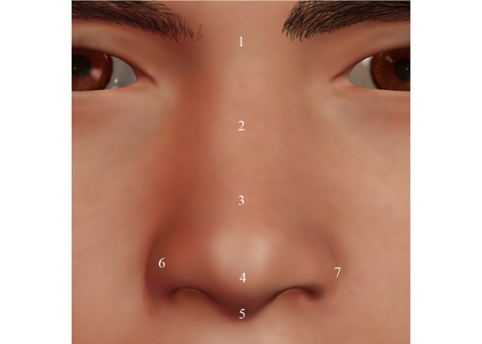 Figure 3. Nasal region: 1 = Nasal root (nasion); 2 = Nasal bridge; 3 = Supratip break; 4 = Nasal tip; 5 = Columella; 6 = Ala; 7 = Alar crease.
- Nasal root (nasion): top of the nose that attaches the nose to the forehead, above the bridge and below the glabella.
- Nasal bridge: nasal vault made up of the proximal bone and the distal cartilage.
- Supratip break: subtle dip above the nasal tip made up of the major alar cartilage over the septal and lateral cartilages.
- Nasal tip: the most anterior point of the nose (pronasale).
- Columella: the bridge of flexible tissue that divides the nasal fossae and is made up of the medial crura of the alar cartilages.
- Nasal alae: they form the lateral part (wing) of the nasal vestibule and are made up of the major and minor alar cartilages and fatty tissue.
- Alar crease: the fold located at the intersection of the nasal ala and the cheek.
Labial region
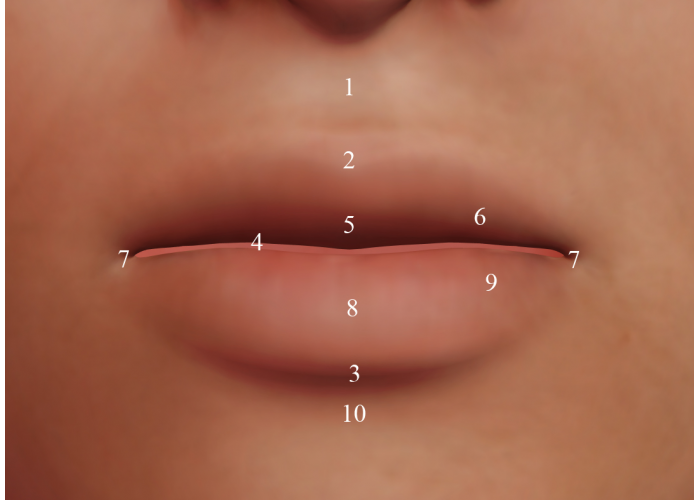 Figure 4. Labial region: 1 = Philtrum; 2 = Upper vermillion border ( cupid’s bow); 3 = Lower vermillion border; 4 = Labial fissure; 5 = Labial tubercle; 6 = Alae; 7 = Commisures (chelion); 8 = Medial sulcus; 9 = Tori; 10 = Mentolabial sulcus.
- Philtrum: vertical medial indentation that extends over the upper lip.
- Upper vermillion border: Sharp demarcation between the upper lip and the adjacent skin; when a coincidence of the peaks of the bow with the philtral columns is present, it may be referred to as “Cupid’s bow”.
- Lower vermillion border: Sharp demarcation between the lower lip and the adjacent skin.
- Labial fissure: the slit between the lips, opening to the oral cavity.
- Labial tubercle: the slightly projected mass of soft tissue on the free edge of the centre of the upper lip. Also referred to as prochelion.
- Alae: the paired wings of the upper lip on each side of the tuber.
- Labial corner: the corner (angle) of the mouth. More commonly known as the cephalometric chelion.
- Medial sulcus: subtle sulcus of the midline on the lower lip. el ligero surco de la línea media en el labio inferior.
- Tori: the paired tubercles that make up the body of the lower lip.
- Mentolabial sulcus: the groove that separates the lower lip from the upper part of the chin protrusion.
Auditory region
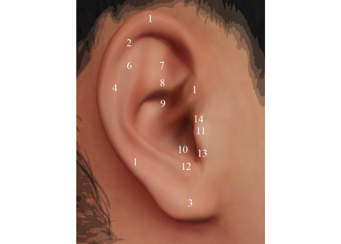 Figure 5. Auditory region: 1 = Helix; 2 = Darwin’s tubercle; 3 = Lobe; 4 = Scapha; 5 = Antihelix; 6 = Superior crus; 7 = Triangular fossa; 8 = Inferior crus; 9 = Concha cymba; 10 = Concha cavum; 11 = Tragus; 12 = Antitragus; 13 = Intertragal notch; 14 = Tragion.
- Helix: the outer rim of the ear (external).
- Darwin’s tubercle: variable congenital ear condition that manifests as a thickening of the helix towards the upper portion of the helix.
- Lobe: the fleshy part that hangs from the lower margin of the ear.
- Scapha: the groove between the helix and the antihelix.
- Antihelix: the “Y” shaped crest in the interior of the auricula.
- Superior crus: the upper cartilaginous ridge of the antihelix.
- Triangular fossa: the concavity between the two ends (crura) of the antihelix.
- Inferior crus: the lower cartilaginous ridge of the antihelix.
- Concha cymba: the fossa bounded by the tragus, antitragus, antihelix and the inferior crus of the antihelix.
- Intertragal canal/Concha cavum: the fossa that connects to the concha cymba and is located right under the root of the helix, leading to the external auditory canal.
- Tragus: cartilaginous prominence in the inner side of the external ear, right in front of the opening of the external ear canal.
- Antitragus: cartilaginous protuberance in the lower margin of the antihelix, opposed to the tragus.
- Intertragal notch: the groove between the tragus and the antitragus.
- Tragion: cephalometric landmark located at the conjuncture of the helix and the tragus, lying on top of its craniometric homologue.